

[Jan-2025] RhMSUS Free Sample Questions to Practice One Year Update
Download RhMSUS exam with American College of Rheumatology RhMSUS Real Exam Questions
NEW QUESTION # 41
Which of the following anatomic structures BEST fits the scope of rheumatologic ultrasound practice?
- A. Posterior patellar cartilage
- B. Metacarpal volar plate
- C. Posterior cruciate ligament
- D. Anterior glenoid labrum
Answer: A
NEW QUESTION # 42
Ultrasound evaluation of a painful shoulder is MOST likely to be useful in planning a corticosteroid injection for which of the following conditions?
- A. Superior labral anterior-posterior lesion
- B. Adhesive capsulitis
- C. Complex regional pain syndrome
- D. Glenohumeral synovitis
Answer: D
NEW QUESTION # 43
This is a short-axis scan of the supraspinatus tendon in a normal person. What is the finding marked with the arrow?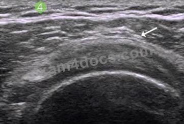
- A. Calcification of the supraspinatus tendon
- B. Subacromial bursa calcification
- C. A probe-generated artifact
- D. Normal deltoid muscle
Answer: D
NEW QUESTION # 44
A practitioner wants to assess the structures on the right, distal portion of this image.
What should the practitioner do at this time?
- A. Choose a lower transducer frequency.
- B. Place the focal point at the level of the area of interest.
- C. Increase the overall gain to brighten the image.
- D. Depress the distal end of the probe.
Answer: B
NEW QUESTION # 45
Which of the following protocols would be MOST consistent with a complete routine ultrasound examination of the shoulder?
- A. Anterior long- and short-axis views of the biceps tendon
oAnterior view of the rotator cuff interval
oLateral views of the subscapularis tendon
oLateral long- and short-axis views of the supraspinatus tendon, moving the transducer anteriorly and posteriorly oPosterior view of the glenohumeral joint recess oDynamic lateral long-axis assessment of impingement with abduction of the arm - B. Anterior long- and short-axis views of the biceps tendon
oAnterior views of the pectoralis tendon insertion
oAnterior view of the coracoid ligament and rotator cuff interval
oLong- and short-axis views of the supraspinatus tendon, moving the transducer anteriorly and posteriorly oPosterior long- and short-axis views of the infraspinatus tendon insertion oAxillary long-axis view with arm raised - C. Anterior long- and short-axis views of the biceps tendon
oAnterior view of the subscapularis tendon in external rotation
oLong- and short-axis views of the supraspinatus tendon, moving the transducer anteriorly and posteriorly oPosterolateral view of the infraspinatus tendon oPosterior view of the glenohumeral joint recess oView from cranial at the acromioclavicular joint - D. Anterior long- and short-axis views of the biceps tendon
oAnterior view of the rotator cuff interval
oLong- and short-axis views of the supraspinatus tendon, moving the transducer anteriorly and posteriorly oDynamic lateral long-axis assessment of impingement with abduction of the arm oPosterior transverse view of the suprascapular notch oView from cranial at the acromioclavicular joint
Answer: C
NEW QUESTION # 46
At approximately how many degrees of flexion is the MOST sensitive resting position of the knee for imaging fluid in the suprapatellar recess?
- A. 0
- B. 1
- C. 2
- D. 3
Answer: A
NEW QUESTION # 47
What is the MOST prominent ultrasound artifact depicted in this image?
- A. Enhancement
- B. Comet tail
- C. Ring down
- D. Shadowing
Answer: A
NEW QUESTION # 48
Which condition is the ultrasound examination of this nodular structure MOST suggestive of?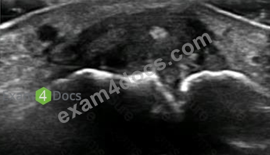

- A. Rheumatoid nodule
- B. Gouty tophus
- C. Ganglion cyst
- D. Subcutaneous abscess
Answer: C
NEW QUESTION # 49
Which of the following is the MOST appropriate next step to confirm the presence of pathology in this metacarpophalangeal joint?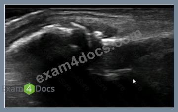
- A. Decrease the depth.
- B. Check the transverse plane.
- C. Float the probe.
- D. Use a "hockey stick" probe.
Answer: D
NEW QUESTION # 50
An 81-year-old male with known shoulder osteoarthritis presents to the office for an ultrasound-guided injection of the glenohumeral joint. Prior to injection, where should the needle tip be visualized?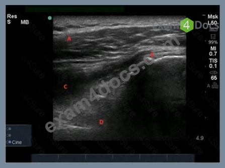
- A. A
- B. B
- C. D
- D. C
Answer: C
NEW QUESTION # 51
The image below shows the injection of the dorsal wrist region in a patient with rheumatoid arthritis. The needle is indicated by the arrows. Considering the ultrasound findings, which of the following is MOST appropriate in completing the injection?
- A. Both the radiocarpal and midcarpal recesses should be targeted.
- B. The fourth compartment ten synovium should be targeted.
- C. The midcarpal recess should be the primary target.
- D. The radiocarpal recess should be the primary target.
Answer: A
NEW QUESTION # 52
This posterior transverse scan of the shoulder of a 3-year-old boy shows which of the following?
- A. Normal shoulder scan
- B. Effusion with an erosion of the humeral head
- C. Effusion of the glenohumeral joint
- D. Fracture of the humeral head
Answer: C
NEW QUESTION # 53
What is the position of the arm MOST likely used to obtain this B-Mode image of the shoulder?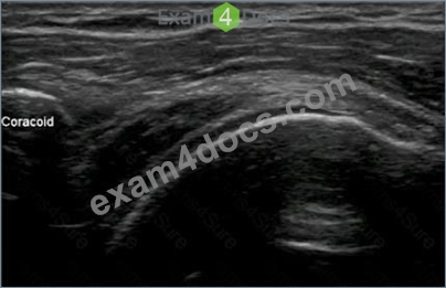
- A. External rotation
- B. Crass
- C. Neer impingement
- D. Modified Crass
Answer: D
NEW QUESTION # 54
What should be taken into consideration prior to any procedure?
- A. The shortest pathway to the region of interest should be selected with consideration given to adjacent neurovascular structures.
- B. A slight to-and-fro movement or injection of a small amount of lidocaine may be beneficial in visualizing the needle.
- C. The needle should be visualized only prior to its insertion to the target area.
- D. The transducer should be aligned perpendicular to the plane of the needle.
Answer: A
NEW QUESTION # 55
For which of the following clinical scenarios is it appropriate for a practitioner to perform musculoskeletal ultrasound?
- A. A patient with psoriasis and posterior heel pain
- B. A patient who has pain and numbness over the lateral calf
- C. A 40-year-old man with a snapping sensation in his groin with hip flexion and external rotation
- D. A 50-year-old woman with pain between the third and fourth metatarsals
Answer: C
NEW QUESTION # 56
A 62-year-old man presents with left shoulder pain. The ultrasound image was taken at the patient's maximum abduction (45 degrees). Which of the following rotator cuff pathologies is BEST evaluated with this shoulder position?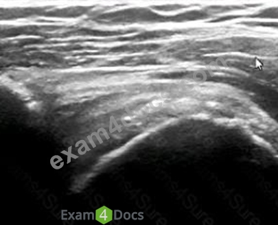
- A. Complete tear
- B. Enthesopathy
- C. Intrasubstance tear
- D. Impingement
Answer: D
NEW QUESTION # 57
A 3-year-old child with juvenile idiopathic arthritis presents for an intra-articular corticosteroid injection of the knee. Using ultrasound guidance, which location should be targeted with a needle?
- A. A
- B. B
- C. D
- D. C
Answer: D
NEW QUESTION # 58
A 30-year-old pregnant woman with rheumatoid arthritis presents with right wrist pain and swelling despite a palpation-guided corticosteroid injection one month ago.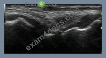
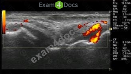
- A. Extensor tenosynovium
- B. Radioulnar joint
- C. Intercarpal joint
- D. Radiocarpal joint
Answer: C
NEW QUESTION # 59
How is the dynamic evaluation for subacromial impingement and adhesive capsulitis performed?
- A. By placing the probe in a coronal-oblique position over the distal clavicle and asking the patient to raise the arm
- B. By placing the probe in a transverse position over the distal clavicle and asking the patient to raise the arm
- C. By placing the probe in a longitudinal position between the greater tuberosity and acromion and asking the patient to raise the arm.
- D. By placing the probe in a transverse position between the greater tuberosity and acromion and asking the patient to raise the arm
Answer: C
NEW QUESTION # 60
By comparing gray-scale images from before pharmacologic treatment and after months of treatment, rheumatologists are able to assess patient improvement and response with regard to:
- A. Articular cartilage damage in osteoarthritis.
- B. Erosions in rheumatoid arthritis.
- C. Osteophytes in osteoarthritis.
- D. Synovitis in rheumatoid arthritis.
Answer: D
NEW QUESTION # 61
What is the BEST description for this longitudinal ultrasound scan of a 75-year-old patient with pain in the medial aspect of the knee?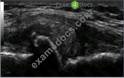
- A. Osteophytes, distension of the medial collateral ligament, and meniscal degeneration
- B. Osteophytes and distension and calcification of the medial collateral ligament
- C. Osteophytes and rupture of the medial collateral ligament
- D. Osteophytes and meniscal cyst
Answer: A
NEW QUESTION # 62
......
Real exam questions are provided for ACR-MUCR tests, which can make sure you 100% pass: https://www.exam4docs.com/RhMSUS-study-questions.html
RhMSUS Exam with Guarantee Updated 100 Questions: https://drive.google.com/open?id=1c5-p4VUWFUZTy1eGetPjm5yfyG70Llgw
